The Tiger's Mouth Blog: Notes on Anatomy and Physiology: More On The Ties That Bind
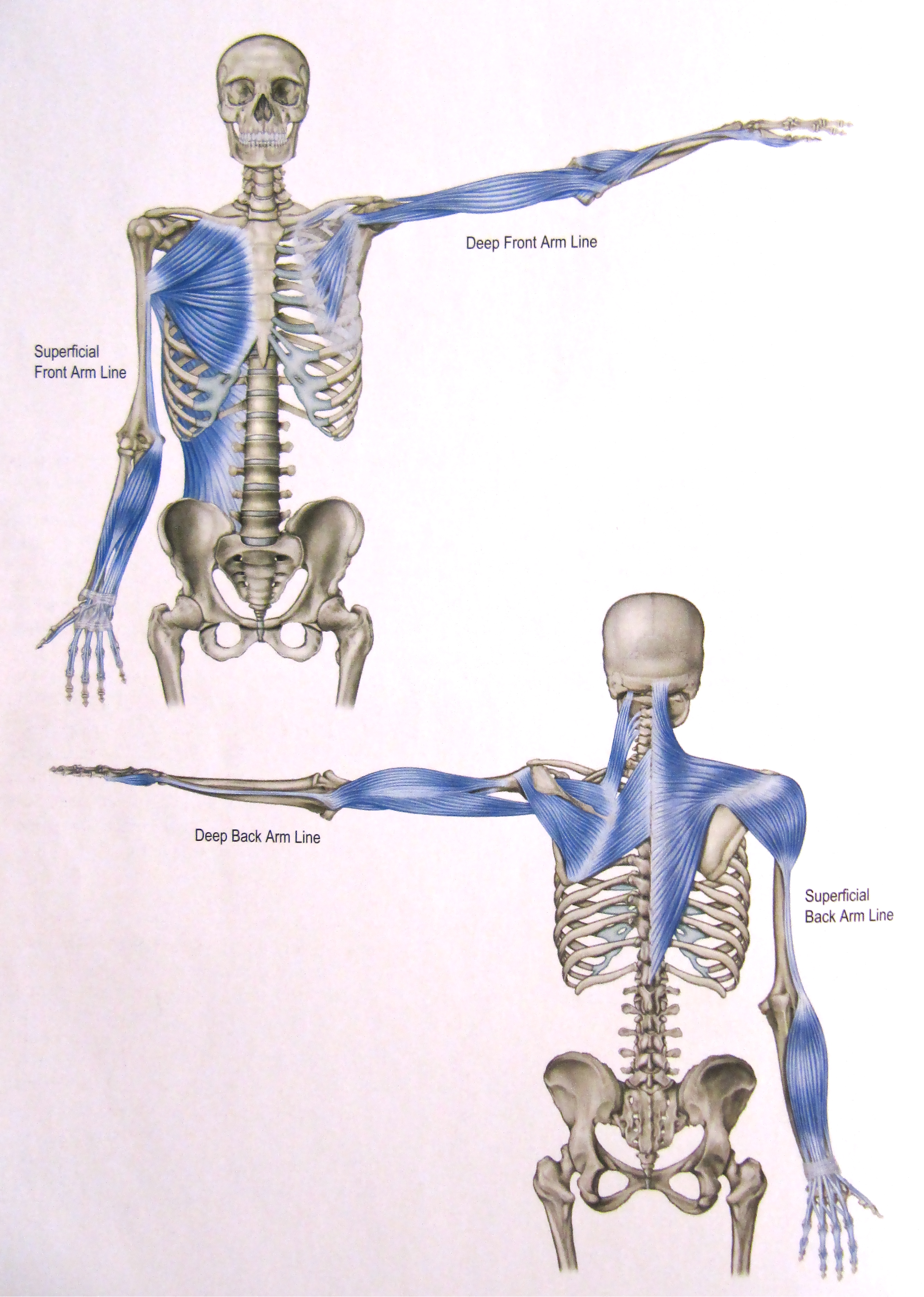
Thomas Myers in his book Anatomy Trains describes four lines of pull that arise, shift and interact in the upper limb as we move.
Two of these lines run along the front of the arm and two others along the back. As you peruse the drawings, look not for detail but for a sense of how these pathways take the upper limb and hook it up with the torso as we move through the set.
Some observations about these lines
- they are labelled superficial or deep depending on whether they attach to surface or deep tissues of the chest wall
- they each travel from the axial skeleton out to one of the four sides of the hand – thumb, little finger, palm or back of the hand. Or, they could be described as running from the four sides of the hand in to the axial skeleton. These lines of influence traverse the body in both directions.
- part of the power of Tiger’s Mouth arises from its ability to simultaneously engage all aspects of the hand and act as an endpoint for each one of these lines
- the Superficial Front Arm Line connects the front of all five digits and the palm to two large muscles on the chest wall: the pectoralis major at the front and the latissimus dorsi (not visible in the drawing) at the back
- the Deep Front Arm Line runs from the thumb up through the biceps and onto the pectoralis minor
- the Superficial Back Arm Line joins the back of the hand with the shoulder blade, spine and skull
- the Deep Back Arm Line flows from the little finger to the triceps, shoulder blade, spine and skull
- by means of these pathways, the hand hitches up with substantial central structures that have their own extensive connections, thus amplifying the breadth of interaction.
Let’s look at three of the central structures with which the hands connect: the pectoralis major, the latissimus dorsi and the pectoralis minor. Pectoralis Major Broad and powerful, this muscle twines about itself as it runs from the medial half of the collar bone, the sternum and the cartilage of the first 6 ribs over to the upper humerus, eventually connecting with the front of the hand by a transmission line of muscle, tendon, fascia and bone. Functions of the muscle itself include drawing the arm in toward the side (adduction) and the front of the body (flexion).
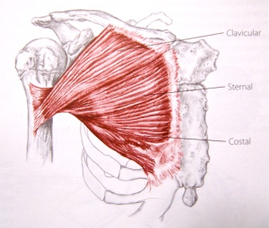
This next image brings to mind the fourth arm foundation exercise, and the links that form between the two hands as we rise up and open in Brush Knees.

Latissimus Dorsi The latissimus dorsi has extensive proximal attachments with the torso: the spinous processes of the sixth thoracic to fifth lumbar vertebrae (T6-L5), the sacrum, the posterior iliac crest, the thoracolumbar fascia, the lower 4 ribs and the inferior tip of the shoulder blade. Spiralling as it approaches the arm, the latissimus grabs hold of the anterior upper humerus right beside the insertion of the pectoralis major. Like the pectoralis major, it too eventually links up with the front of the hand. Among other things, this muscle serves to draw the arm in and back behind the body.
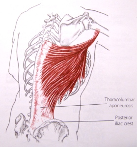
Because the hand eventually makes fast to both the pectoralis major and the latissimus dorsi, it can connect powerfully with the torso.

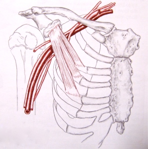
Pectoralis minor The pectoralis minor sits on the rib cage, deep to the pectoralis major. It runs from the 3rd, 4th and 5th ribs up to the coracoid process, an anterior projection of the scapula used to attach ligaments and muscles. As a result, this muscle contributes to the already very mobile life of the shoulder blade by drawing it laterally along the posterior chest wall and tilting it anteriorly. As we saw in the Deep Front Arm Line, the pectoralis minor ties the thumb to the front of the chest.
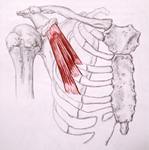
The artery, vein and nerves serving the upper limb pass underneath the pectoralis minor. Our keeping this muscle supple, preventing it from shortening and crowding the space beneath, helps avoid compression of the neurovascular bundle as it makes its way into the arm.
So, multiple channels of potential connection run from the upper limb to various regions of the chest, scapula, spine and head. Using our hands, wrists and elbows with the right intention activates these various pathways in a manner that contributes strength, balance and ease to our movements. Too little intention and connection is not established. Too much and the body tenses; we lock into one line of pull and find it difficult to shift into others.
Next time, we will see how the whole body continuities are completed as the torso joins with the pelvis and lower limbs. Hands linked to trunk, head and feet.
1. Kinesiology of the Musculoskeletal System, Foundations for Rehabilitation, 2nd Edition, 2010, Donald A. Neumann, Mosby Elsevier, ISBN 978-0-323-03989-5
2. Trail Guide to the Body, 3rd Edition, 2005, Andrew Biel, Books of Discovery, ISBN 0-9658534-5-4
