Blogue de la Bouche du tigre: Notes on Anatomy and Physiology: The Spinal Ligaments – Holding All The Parts Together
Thus far we have been introduced to a number of the components of the spinal column – the vertebrae themselves, the intervertebral discs and the facet joints. We have looked briefly at both lumbar disc disease and spinal stenosis and gained a sense of how these conditions affect, and are affected, by posture and movement in quite different ways.
We’ve seen, as well, that the intervertebral discs and facet joints provide the spine with stability as they tie the 24 vertebrae together into a coherent whole. While at the same time bestowing mobility and permitting each vertebra to move on its neighbours. 24 articulations sitting one on top of the other and connecting skull to pelvis.
The spinal ligaments work in much the same manner. They play a crucial role in preventing the spine from collapsing into a pile of bones at our feet. But they also allow movement within a predetermined range, winding up like springs in the process and storing elastic energy that can then be used to return the spine to its previous posture.
As we describe the elements of this ligamentous system, keep in mind that they all come together to form a continuous, dense connective tissue stocking around the spine – a stocking that then reaches out to the entire body, connecting the vertebral column and its contents with the torso, head, limbs and sacrum. And remember that we play constantly with these ligaments as we practice the Taoist Tai Chi® arts.

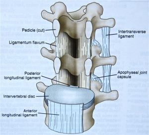
The spinal ligaments consist of:
- The anterior longitudinal ligament that runs from the base of the skull along the front of each vertebral body and disc and down the anterior sacrum. It fuses with a sheath, the periosteum, that wraps tightly around each vertebra. This ligament is the only spinal ligament that resists backward bending and limits the forward curve of the neck and lumbar regions.
- The posterior longitudinal ligament that also melds with the periosteum of the vertebral bodies and skull but this time posteriorly. It runs along the back aspect of the vertebral bodies and discs and down into the canal that lies within the sacrum. It offers attachment sites for the dural sac of the spinal canal. It tightens with forward bending.
- The facet joint capsule, a balloon like structure that wraps around each facet joint. Its sensory receptors likely guide the movement between adjacent vertebrae.
- The ligamentum flavum that connects the back of the vertebral arches and forms the back wall of the spinal canal. It is known as the yellow ligament because of the colour imparted by the preponderance of elastic fibers. Off to the sides, it fuses with the facet joint capsules. In the midline, it turns posteriorly to become the interspinous ligament. Lengthened by flexion (forward bending) of the spine, its elastic fibers supply a strong returning force. The area of the spine with the most flexion, the lumbar region, is where the ligamentum flavum is the thickest.
- The interspinous ligament running between the spinous processes. Its more anterior fibers are rich in elastin and blend with the ligamentum flavum; the more posterior fibers meld with the supraspinous ligament.
- The intertransverse ligaments that bind the ends of the transverse processes and resist side bending to the opposite side.
- The supraspinous ligament that connects the tips of the spinous processes and goes on to meld with the thoracolumbar fascia, a sheet we will describe in the next note.


These spinal ligaments, the discs and the facet joint capsules are all connective tissue structures that need to maintain their strength and flexibility if they are to continue to both brace the spine and allow it mobility. Regular, intermittent straightening of the spinal curves, rotation of the vertebrae through their physiologic range of motion and slight separation of each pair of vertebrae along the full length of the spine as the body expands and contracts, all serve to delay the aging of this connective tissue that comes with time, injury and inactivity. The don yu and tor yu are beautifully designed to accomplish this.
Next time, we’ll look at another vital structure of the spine that is richly engaged in the practice hall – the thoracolumbar fascia.
1. Atlas of Human Anatomy, 4th Edition, 2006, Frank H. Netter, Saunders Elsevier, ISBN-13: 978-1-4160-3385-1
2. The Physiology of the Joints, Volume Three, The Spinal Column, Pelvic Girdle and Head, AI Kapandji, Sixth Edition, 2008, Churchill Livingston, ISBN-10: 0702029599

